
RIS vs PACS: The Complete 2025 Guide
Learn how Radiology Information Systems (RIS) & Picture Archiving and Communication Systems (PACS) work together in our 2025 radiology systems guide.
16 min read

Medical images pile up fast. One busy imaging department can create thousands of X-rays, scans, and ultrasounds every week. For decades, all of it lived on physical film.
Think about what that used to mean. Someone had to develop each film, file it, carry it down the hall, and hope it never went missing. When a doctor needed a scan from last year, the search could take hours.
It sounds slow. It was.
A PACS system fixes that. Short for Picture Archiving and Communication System, it is the digital backbone that captures, stores, shares, and shows medical images across a facility. Instead of hunting for film, a doctor can pull up an image seconds after it is taken.
This guide walks you through what a PACS system is, how it works, and why it matters. You will learn what the letters stand for, the main jobs it handles, and the four parts that make it run. We will also cover the real benefits for your team and how it connects to your other systems.
Whether you run an imaging department or just want to understand the tools behind modern scans, this is your plain-language start. No dense jargon. No filler. Just a clear look at the technology that quietly powers nearly every scan a patient gets today.
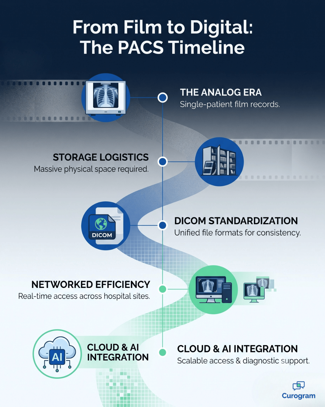
A PACS system is the digital heart of a modern imaging department. It changed how images are handled from the ground up.
Before it arrived, imaging departments were full of physical film and bulky storage rooms. Sharing images was slow and done by hand. The whole point of a PACS system is to swap those old methods for a digital one. It captures, stores, shares, and shows medical images on a screen.
Here is what that looks like in practice.
A doctor can view an X-ray or MRI moments after the scan. It does not matter where in the hospital the patient was imaged.
In some cases, they can even do it from home.
This does more than speed up reads. It helps teams work together, since specialists can view the same images at the same time from different places. So when someone asks what is PACS in radiology, the honest answer is a leap toward faster, clearer, more connected care.
Many wonder, what exactly does PACS stand for? The term PACS stands for Picture Archiving and Communication System.
Each word in this acronym highlights a critical function of this technology in a PACS system radiology environment.
Put those four words back together and you get the full picture. A PACS system is a connected setup built to handle images from the moment they are captured to the day they are pulled up years later.
A PACS medical system does far more than store pictures. It manages the whole life of an image, from capture to final report. Each job below works with the others to keep scans secure, sorted, and ready when your team needs them.
The journey of a medical image within a PACS radiology environment begins with acquisition and import into the PACS system. Image acquisition involves capturing images directly from various medical imaging devices, known as modalities.
These include:
Computed Radiography (CR),
Digital Radiography (DR),
Computed Tomography (CT),
Magnetic Resonance Imaging (MRI),
Ultrasound (US), and others.
Once an image is captured, it is typically converted into the DICOM (Digital Imaging and Communications in Medicine) format. The DICOM standard is globally recognized and ensures that medical images and related data are standardized for handling, storing, printing, and transmitting information in medical imaging.
This standardization is fundamental for interoperability within the PACS system and between different PACS medical systems or related healthcare IT systems.
The PACS system then ingests these DICOM files, associating them with patient demographic information, usually retrieved from a Radiology Information System (RIS) or Hospital Information System (HIS).
Secure archiving and robust storage are foundational functions of any PACS system, critical for maintaining patient data integrity and ensuring long-term accessibility in a pacs medical setting. Once images are acquired, the PACS system archives them in a structured digital library. This often involves a tiered storage architecture.
For instance, recent or frequently accessed images might be stored on faster, more expensive storage like a Redundant Array of Independent Disks (RAID) for quick retrieval. Older, less frequently accessed images might be moved to more cost-effective, long-term archive solutions, including cloud storage or magnetic tape libraries.
Data security is paramount:
The PACS system must comply with regulations like HIPAA (Health Insurance Portability and Accountability Act) in the United States, ensuring patient confidentiality through encryption, access controls, and audit trails.
Disaster recovery plans are also integral to the archiving strategy of a pacs system radiology department, ensuring that images can be recovered in case of hardware failure, natural disaster, or cyber-attack.
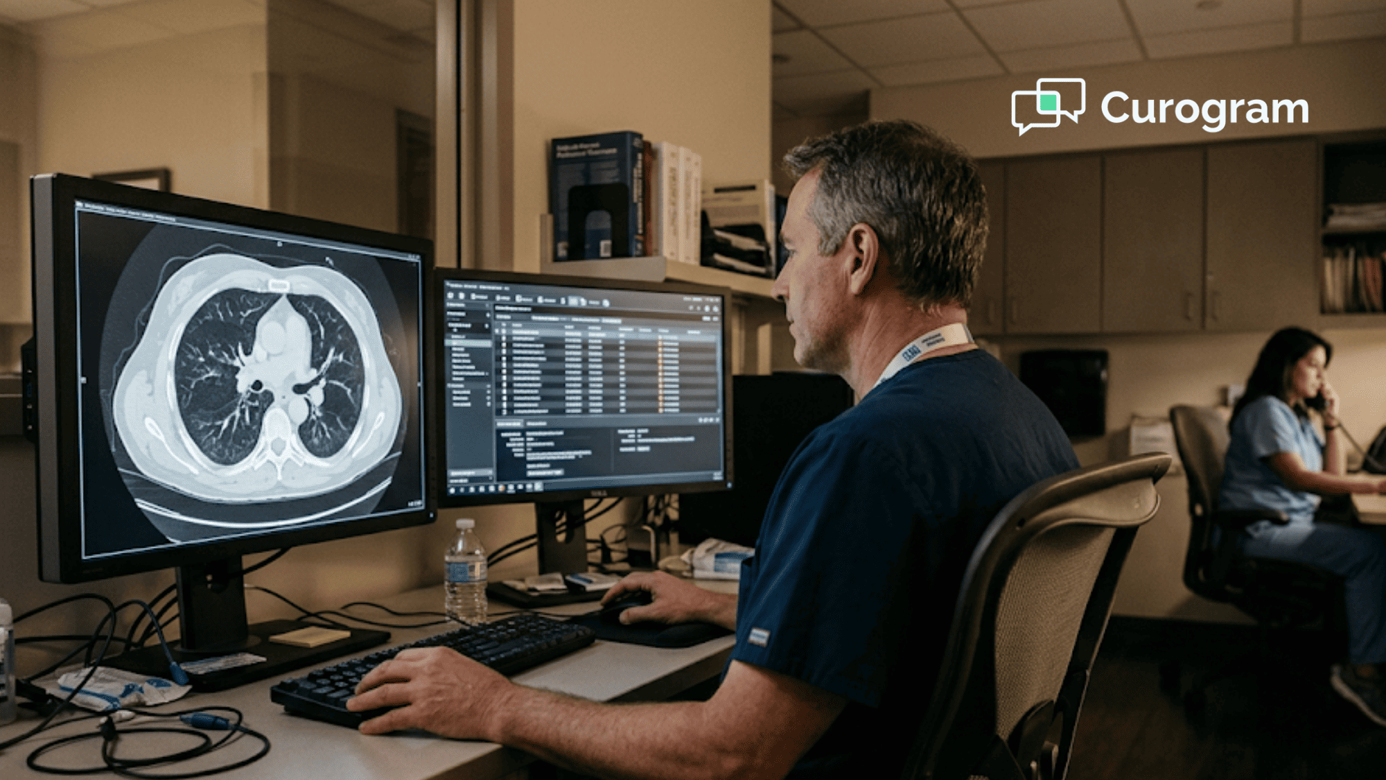
The ability to effectively display and manipulate medical images is a cornerstone of the diagnostic process, heavily reliant on sophisticated PACS software within the PACS system. Radiologists and other clinicians use specialized viewing workstations equipped with high-resolution medical-grade monitors to examine images.
The PACS software provides a rich set of tools for image manipulation, far exceeding the capabilities of traditional film. These tools allow users to zoom, pan, magnify specific areas, adjust brightness and contrast, invert grayscale, annotate images with text or measurements, and compare current images with prior studies side-by-side.
Advanced PACS software may also include 3D reconstruction capabilities, allowing for volumetric viewing of datasets (e.g., from CT or MRI scans), providing deeper insights for diagnosis and surgical planning.
The quality and functionality of the PACS software directly impact the diagnostic efficiency and accuracy within the pacs medical environment.
Efficient image retrieval and distribution are key benefits of a PACS system radiology departments leverage to speed up patient care. Unlike the cumbersome process of physically finding and transporting film jackets, a PACS system allows authorized users to retrieve patient images almost instantaneously from the digital archive.
Using search criteria like patient name, ID, date of study, or modality, clinicians can quickly access the required images on their workstations. Distribution is also greatly enhanced. Images can be electronically sent to referring physicians' offices, specialist consultation rooms, surgical theaters, or even to other hospitals if a patient is transferred.
Many PACS system setups include web-based viewers, which allow secure access to images from standard computers or mobile devices, further broadening accessibility for authorized personnel.
This rapid retrieval and distribution capability within a pacs medical setting is crucial for timely diagnosis, emergency care, and collaborative treatment planning.
A modern PACS system also keeps the department running smoothly. Tied closely to the RIS, it helps manage the full imaging flow.
Smart worklists sort scans by urgency, so an emergency case jumps to the front. The system tracks each scan through its stages, from scheduled to read to reported. That trims the wait for results. Add voice-to-text for reports, and doctors spend less time on manual steps and more time on care.
|
Quick takeaway: A PACS system is not just a filing cabinet for scans. It captures, protects, shows, moves, and tracks every image. So your team can find the right scan in seconds instead of searching for a missing film for an hour. |
To truly understand what is PACS in radiology, it's essential to recognize the four major components that constitute a functional PACS system. These components work in concert to deliver the efficiency and benefits expected from a modern pacs medical imaging solution.
Each part of the PACS system plays a distinct role, from image creation to its final interpretation and storage, forming the technological backbone of any pacs system radiology department.
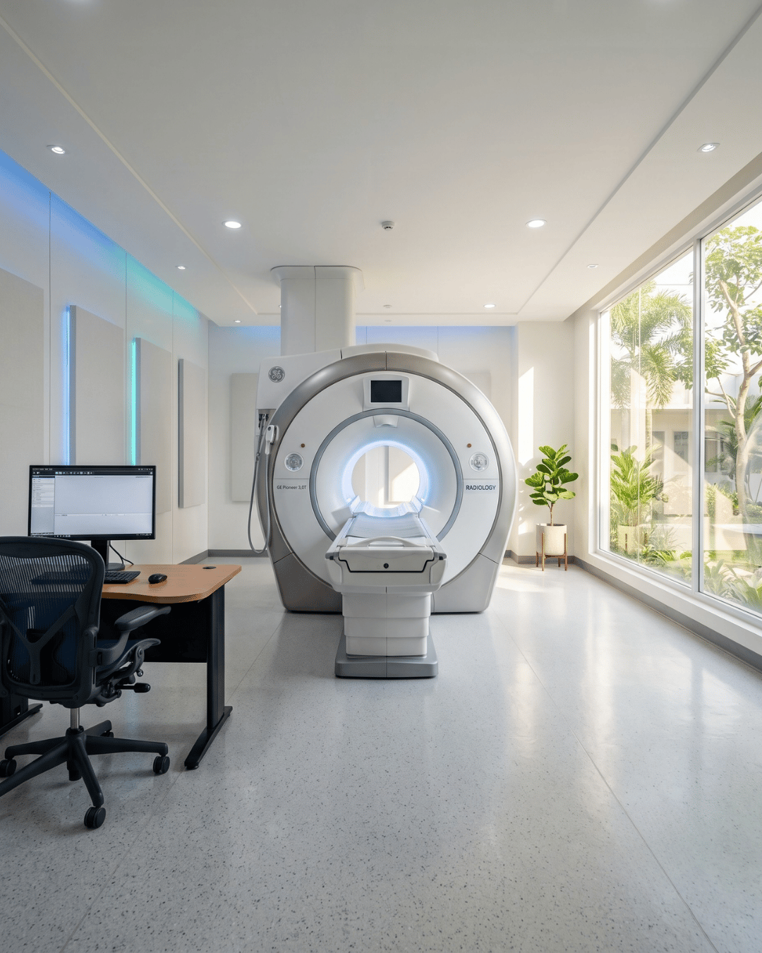
Imaging modalities are the various types of sophisticated medical equipment that actually capture the patient images, forming the initial input source for the PACS medical data pipeline within a PACS system.
These are the machines at the frontline of diagnostic imaging. Examples include:
Each machine sends digital image data, usually as DICOM files, to the central system. The quality of that data shapes everything that follows.
A secure and robust network is the communication backbone that enables the "C" (Communication) in PACS, absolutely critical for any PACS radiology department.
This network connects all the other components of the PACS system, allowing vast amounts of image data to flow between imaging modalities, storage archives, display workstations, and other integrated systems like the RIS or EMR.
Given the large file sizes of medical images (especially from CT, MRI, and digital mammography), the network infrastructure, including switches, routers, and cabling (often high-speed fiber optic), must have sufficient bandwidth to handle the traffic without causing bottlenecks.
Slow network performance can severely hamper the efficiency of a pacs system radiology department, leading to delays in image access and diagnosis.
Furthermore, because pacs medical data is sensitive patient information, the network must be highly secure, incorporating firewalls, intrusion detection systems, and secure protocols (like VPNs for remote access) to protect against unauthorized access and cyber threats, ensuring the integrity of the PACS system.
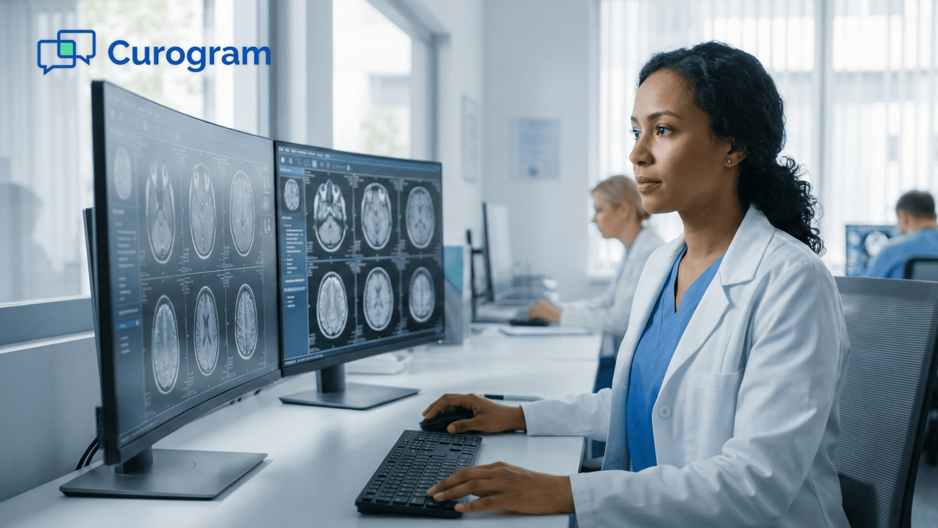
PACS workstations are the specialized, high-performance computer systems where radiologists and other clinicians view, interpret, and report on medical images, effectively serving as the command center within the PACS system.
These workstations are equipped with one or more high-resolution, medical-grade monitors specifically designed to display grayscale images with the precision needed for accurate diagnosis.
The monitors used in pacs radiology are often much higher quality than standard consumer displays to show subtle details.
The workstations run sophisticated PACS software that provides tools for image manipulation (zoom, pan, window/level adjustments, measurements, MPR/3D reconstructions), comparison with prior studies, and often integration with reporting systems, sometimes using voice recognition.
The ergonomic design of the workstation area is also important for the comfort and efficiency of radiologists who spend many hours interpreting images.
The performance and features of these workstations directly influence the diagnostic capabilities and workflow efficiency of the pacs medical team using the PACS system.
Archives and servers form the very heart of the PACS system, responsible for the long-term storage, management, and retrieval of all medical images and associated data within the pacs radiology ecosystem.
The core of this component is usually a powerful server running a database that catalogs all the images and patient information.
The actual image storage can involve various technologies:
Backups matter a lot here, since losing images is not an option. The archive also has to grow, because scan volumes only rise.
PACS software is the intelligent engine that drives the functionality and usability of the entire PACS system, transforming a collection of hardware components into a cohesive and powerful imaging solution.
The quality and features of the PACS software directly determine how effectively users can interact with medical images, manage workflows, and integrate with other healthcare IT systems.
For any radiology pacs deployment, the choice of software is as critical as the hardware it runs on, as it directly impacts diagnostic confidence and departmental efficiency.
There are several categories of PACS software that work together within a comprehensive PACS system. Viewer software, for instance, is what radiologists use daily, providing tools for image display, manipulation (like adjusting contrast or making measurements), and navigating complex patient studies. Archive software manages the storage, retrieval, and security of image data, ensuring images are correctly filed and can be quickly located.
Workflow engine software, often integrated with the RIS, helps to manage study lists, assign cases to radiologists, and track reporting progress.
Key features to look for in modern PACS software include an:
Intuitive user interface (crucial for reducing learning curves and improving radiologist efficiency),
Robust tools for 2D and increasingly 3D/4D image visualization,
Seamless integration with RIS and EMR/EHR systems (to provide a holistic view of patient data), and
Strong support for DICOM standards and other interoperability protocols.
Advanced PACS software may also incorporate AI-powered tools for tasks like image analysis or workflow prioritization. The ability for the PACS software to support web-based and mobile viewers is also increasingly important, allowing for remote consultations and flexible access to images within the radiology pacs environment.
Ultimately, the right PACS software empowers the entire PACS system to enhance patient care.
Implementing a PACS system brings a multitude of transformative benefits to radiology pacs operations and the broader healthcare organization, revolutionizing efficiency, accuracy, and patient care. The shift from analog film to a digital PACS system fundamentally enhances how medical images are captured, viewed, stored, and shared, leading to significant improvements across various aspects of a pacs medical environment.
A primary benefit of a PACS medical imaging system is the potential for enhanced diagnostic accuracy. PACS software provides radiologists with sophisticated tools to manipulate digital images, such as zooming in on suspicious areas, adjusting brightness and contrast to highlight subtle findings, and making precise measurements.
The ability to easily compare current images side-by-side with prior studies stored in the PACS system allows for better tracking of disease progression or response to treatment.
High-resolution medical displays used with pacs radiology workstations also contribute to better image visualization than traditional film. These capabilities collectively empower radiologists to make more confident and accurate diagnoses.
PACS radiology teams experience dramatically streamlined workflows and increased operational efficiency. The PACS system eliminates time-consuming manual processes associated with film, such as developing, transporting, filing, and retrieving film jackets.
Images are available almost instantly on workstations after acquisition.
Digital worklists can be customized and prioritized, ensuring that urgent cases are read promptly.
Integration with RIS and voice recognition systems further accelerates the reporting process.
This overall increase in speed and efficiency within the PACS system means radiologists can read more studies, and report turnaround times are significantly reduced, benefiting both the department and patient care.
The upfront cost can be steep, but the long-term savings usually make up for it. The clearest savings come from cutting film, chemicals, and processor upkeep for good.
Storage costs drop too, since digital archives take up almost no space and need far less handling. Here is a simple example to show the idea. A mid-sized department that once spent about $60,000 a year on film, chemicals, and storage could free up most of that budget after going digital.
| Cost area | Film-based (yearly, example) | Digital PACS (yearly, example) |
|---|---|---|
| Film and chemicals | $35,000 | $0 |
| Physical storage and handling | $15,000 | $2,000 |
| Retrieval and transport labor | $10,000 | $2,500 |
| Rough total | $60,000 | $4,500 |
Treat those numbers as an example, not a quote. Real figures vary by department size and vendor.
For your team, the point is simple. The steady costs of film add up, and much of that spend goes away with a digital system.
Ultimately, a PACS medical environment contributes to improved patient care. Faster access to images and reports means quicker diagnoses and, consequently, more timely initiation of treatment.
The PACS system facilitates better collaboration among healthcare providers;
Images can be easily shared electronically with referring physicians, specialists in other departments, or even other healthcare facilities if needed for consultations or second opinions.
Many PACS system radiology setups offer secure web-based portals allowing authorized clinicians to view images remotely, which is particularly beneficial for multidisciplinary team meetings and providing care in distributed settings.
A digital PACS system radiology setup offers superior data management and security compared to film-based systems. Digital images are less prone to being lost or misfiled.
The PACS system provides a centralized, organized archive with robust search capabilities. Importantly, digital systems allow for better implementation of security measures to protect patient privacy, including access controls, audit trails to track who has viewed images, and encryption.
Furthermore, pacs medical systems facilitate easier implementation of disaster recovery and business continuity plans, ensuring that critical patient imaging data can be protected and restored in the event of a system failure or catastrophe.
A truly effective PACS system doesn’t operate in a vacuum;
Its power is magnified through seamless integration with other critical healthcare information systems, primarily the Radiology Information System (RIS) and the Electronic Medical Record (EMR) or Electronic Health Record (EHR).
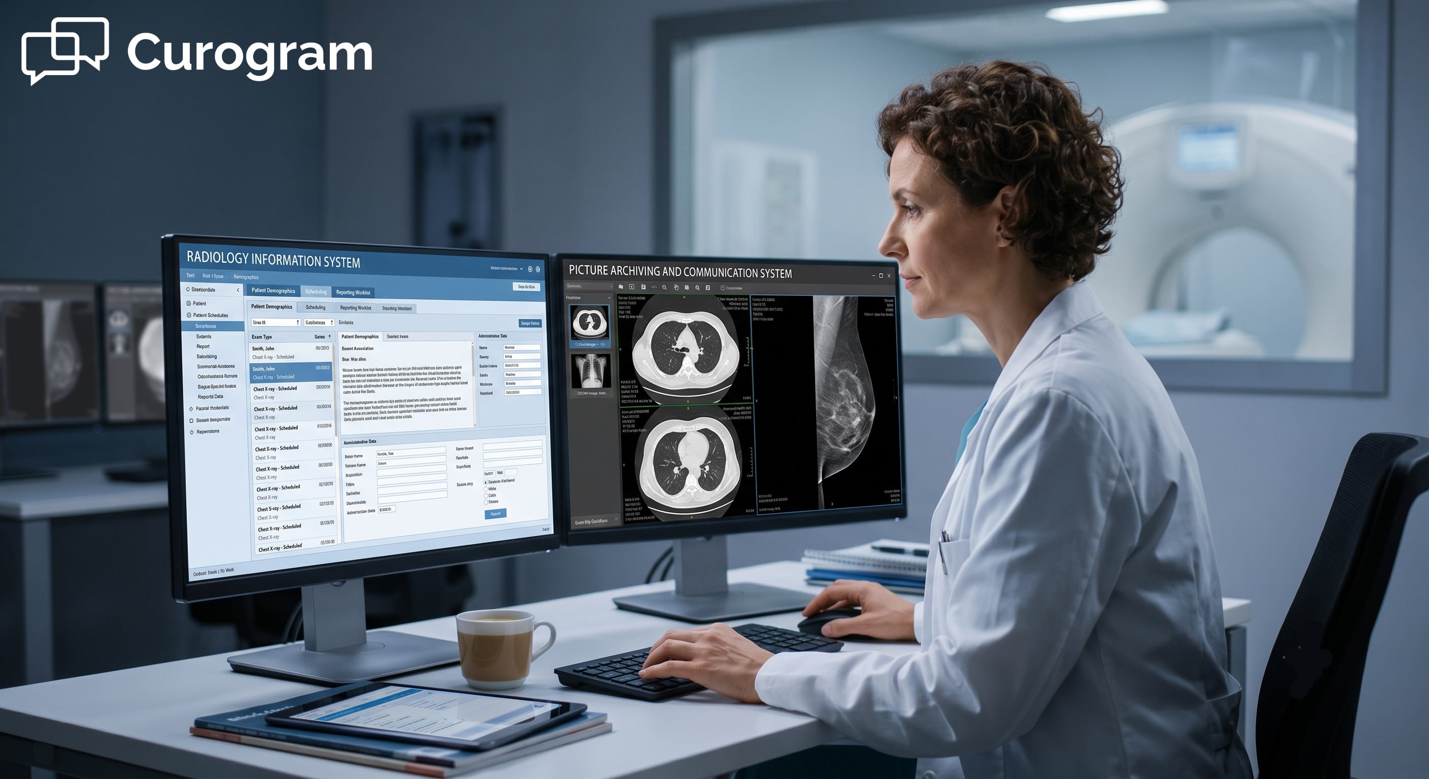
This integration is pivotal for creating a connected pacs medical environment that optimizes workflows and improves data consistency. The RIS typically manages radiology-specific tasks like patient scheduling, exam tracking, and billing.
When integrated with the PACS system, patient demographic data entered into the RIS can automatically populate in the PACS, reducing manual data entry and potential errors.
Similarly, when a radiologist finalizes a report (often dictated using software linked to the PACS), this report can flow back to the RIS and also to the patient's broader EMR/EHR.
Integration with the EMR/EHR allows authorized clinicians across the healthcare enterprise to access not only radiology reports but also the images themselves directly from the patient's comprehensive electronic record. This holistic view of patient information, including imaging from the PACS system, supports better-informed clinical decision-making.
For example,
An oncologist reviewing a patient's progress in the EMR can directly pull up their latest CT scans from the PACS system without needing to log into a separate application.
his level of integration ensures that the valuable data within the PACS medical infrastructure is accessible and utilized effectively across the continuum of care.
The field of PACS radiology is continuously evolving, and one of the most exciting frontiers is the integration of Artificial Intelligence (AI) and Machine Learning (ML).
AI is poised to revolutionize how PACS systems operate and how radiologists interpret medical images.
AI algorithms can be trained to analyze images for subtle patterns that might indicate disease, potentially assisting radiologists by flagging suspicious areas, prioritizing urgent cases, or even performing automated measurements.
This doesn't aim to replace radiologists but to augment their capabilities, improve efficiency, and potentially enhance diagnostic accuracy within the pacs system radiology workflow.
Beyond image analysis, AI can also optimize workflows within the PACS system itself.
For example,
AI could help in protocoling exams, predicting patient no-shows, or managing image archiving strategies more effectively.
Another significant trend shaping the future of pacs medical imaging is the move towards Vendor Neutral Archives (VNAs).
VNAs decouple the image archive from the specific PACS viewing software, giving healthcare organizations more flexibility to switch PACS vendors without complex data migrations.
Cloud-based PACS system solutions are also gaining traction, offering scalability, accessibility, and potentially reduced infrastructure management overhead.
These advancements promise to make PACS radiology even more powerful and integral to patient care.

Selecting and successfully implementing a PACS system is a major undertaking for any healthcare facility, requiring careful planning and consideration of various factors to ensure the chosen pacs medical solution meets current and future needs.
Key considerations include:
Plan the rollout with care and involve everyone who will use it. That means IT, doctors, technologists, and admins. Good training and a clear migration plan make the difference between a smooth switch and a stressful one.
No system is perfect, and a PACS system is no exception. Knowing the common trouble spots up front helps you plan around them.
Here are the ones that trip up practices most often.
Medical images are big, and they pile up quickly. A single CT study can run from 500 MB to over 1 GB.
Picture a department that reads 150 studies a day. At roughly 700 MB each, that is over 100 GB a day, or more than 38 TB a year. In practice, that means storage costs you never planned for can creep up fast.
The fix is simple. Choose a system with room to grow, and lean on tiered or cloud storage so older scans do not clog your fastest drives.
When a PACS system goes down, reads stop. That is the trade-off of going fully digital.
Think about a busy day where your team reads 40 studies an hour.
Even one hour of downtime means 40 delayed reads and 40 patients waiting longer for answers. For your team, that is not just lost time. It is stalled care.
Ask vendors about uptime guarantees, backup servers, and disaster recovery before you sign. A little planning here saves a lot of stress later.
A powerful system helps no one if it does not connect to your other tools or if staff cannot use it. Weak links to the RIS or EMR/EHR push staff back into manual entry. Thin training leads to slow, frustrated users.
Budget time for setup and hands-on training from day one. The teams that adopt fastest are the ones that plan for the learning curve instead of hoping it disappears.
The PACS system has become one of the most essential tools in modern healthcare. It turned medical imaging from a slow, film-based chore into a fast, fully digital flow.
From capturing and storing images to showing them through smart software and linking with your other systems, it supports quicker, sharper reads. It helps clinicians work together. And it leads to better patient outcomes. As AI and cloud tools grow up, the technology will only get more capable and more central to daily care.
Of course, great imaging only helps if the patient actually shows up. Missed CT and MRI slots are costly, and they often come down to a communication gap, not a clinical one. So the patient-facing side of your practice matters just as much as the imaging side.
Curogram's HIPAA-compliant platform helps close that gap.It offers two-way texting, automatic appointment reminders, digital intake forms, and simple patient messaging. Fewer no-shows means your imaging investment gets used to its full worth, and your front desk spends less time on the phone.
If you want to pair a strong imaging setup with smoother patient communication, see how Curogram can support your practice and keep more of those valuable slots filled.
Learn how Radiology Information Systems (RIS) & Picture Archiving and Communication Systems (PACS) work together in our 2025 radiology systems guide.
 Aubreigh Lee Daculug : 2/21/26, 1:00 PM
Understand DICOM imaging standards without technical jargon. Simple explanations with real-world examples for healthcare staff and administrators.
Learn what is RIS in radiology, why it matters to your department, and how it closes the costly gaps between scheduling, reporting, and billing per...